


Diagnostic Imaging
Mason Health offers technologically advanced diagnostic imaging services, including High Field Open MRI and 3D mammography - raising the bar in Mason Health's pursuit of excellence in patient-centered care.
- M-F: 6:30 am to 5:00 pm
- Sat/Sun/Holidays: 9 am to 5:30 pm
MRI, CT Scans, Ultrasounds, General X-Ray
M-F 7:30 a.m. - 6 p.m.
Nuclear Medicine
M-F 7:30 a.m. - 4 p.m.
3D Mammography
M-F 7:30 a.m.- 5 p.m.
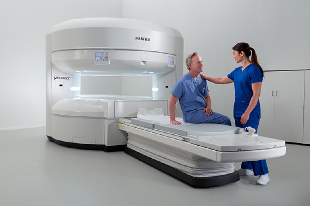
New High Field Open MRI!
The new open MRI will be implemented in the fourth quarter of 2025!
Patients have traveled far and wide for the comfort of Mason Health’s open-field MRI, which the District brought to the community in 2013. Mason General Hospital Foundation is fundraising this year to help Mason Health purchase an upgraded open-field MRI, prioritizing patient comfort and safety. Join us at our next fundraiser! The OASIS Velocity High-Field Open MRI gives providers and technologists an unobstructed view of patients, with an advanced magnet design and imaging capabilities to handle all patients with confidence, without compromising on performance or efficiency. The machine promises fast exams, high-quality images with a seamless workflow.
Some of the MRI’s top attributes include:
- Supporting patients up to 660 pounds on a nearly 3-foot-wide table
- Unique table technology that permits left – right lateral movement, giving patients a more comfortable position
- Excellent image quality using multichannel radiofrequency (RF) technology and Fujfilm RF coil technology
MRI technology combines a powerful magnet, radio waves and a computer to produce high-quality images that assist physicians in assessing a patient’s condition. It differs from both the X-Ray and CT scan because it uses radio waves to pass through the body. This provides exceptional pictures showing soft body tissue in great detail. There are no known side effects. The unit uses no harmful radiation. Nearly any body part may be evaluated from any angle.
New Multifunctional Universal X-Ray Imaging System
Mason Health patients and health care providers will now have a safer and more convenient medical imaging experience for a wide range of procedures, thanks to the purchase of a new state-of-the-art multifunctional universal X-Ray imaging system.
Mason Health’s Shimadzu SONIALVISION G4 LX, made possible by a donation from a grateful patient in March 2022, takes live real-time X-rays for procedures like:
- fluoroscopy
- needle placements
- speech-therapy swallow studies
- gastrointestinal procedures
The imaging system reduces radiation by about 50 percent, compared to previous models, and features a fully articulating bed that can accommodate patients and providers of all heights and abilities.
“The biggest benefits are the dose reduction, the safety, and the diversity of the studies and patient types we can now see,” said Mason Health’s Diagnostic Imaging Manager Shane Faford. “Any bariatric patient we had, we used to have to send them somewhere else. The technology has now changed, and I feel relieved. We not only have state-of-the-art technology, we can use it for multiple purposes, for all the other stuff we need. We’re busier now than we have ever been.”
The imaging system’s bed can lower down to 19 inches from the floor and raise as high as 43 inches, making it easier for patients with limited physical abilities to get in and out of the bed. The bed can also be erected 90 degrees and the arm can extend out 6 feet, making room for patients who need to be seated for a study or who are brought in via wheelchair.
The system is also safer for providers — a protective shield can be raised and lowered for providers who are in the room, and by being able to raise and lower the bed, providers do not have to bend over as much when placing instruments, such as needles, inside of patients. Additionally, the system uses super fine resolution and the most advanced digital imaging processing technology, ensuring the highest quality images.
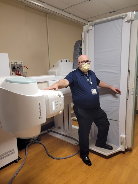
Diagnostic Imaging Manager Shane Faford with the new Shimadzu SONIALVISION G4 LX
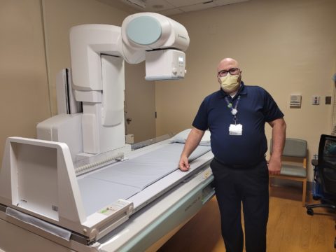
Diagnostic Imaging Manager Shane Faford with the new Shimadzu SONIALVISION G4 LX
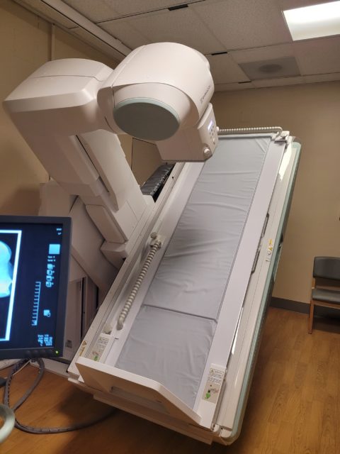
The imaging system’s bed can lower down to 19 inches from the floor and raise as high as 43 inches, making it easier for patients with limited physical abilities to get in and out of the bed. The bed can also be erected 90 degrees and the arm can extend out 6 feet, making room for patients who need to be seated for a study or who are brought in via wheelchair.
Mason Health Now Offers 3D Mammograms for Patients
New 3D Mammography Services Now Available at Mason Health Diagnostic Imaging Department.
Patient Benefits of the Selenia Dimensions 3D Mammography System:
- Ergonomic design for patient comfort
- Up to 40% reduction in false positive recalls, minimizing patient anxiety
- Proven 41% increase in detection of invasive breast cancer
- FDA approved and superior 2D Mammography
Call today to get your mammogram!
3D Mammogram Overview and Video Introduction - Produced by Mason WebTV
Preparing for your MRI
Your MRI requires very little effort on your part. It's a simple process, but preparation is essential! Here's what you need to know:
WHEN YOUR APPOINTMENT IS SCHEDULED
Before we schedule you for an MRI exam, we will ask a number of questions. Your answers will help us identify any current conditions that may make it unsafe for your MRI. You will need to inform us if:
- you are pregnant
- you've ever had surgery
- you're on pain medication
- you're claustrophobic
- you have an incident of metal to your eyes
- you have cardiac valve replacements or a pacemaker, bone or joint replacements, or aneurysm clips, or cardiac stents
These conditions may not exclude you from having an MRI scan, but they'll be important determining factors. Please be prepared to provide information on your insurance and medical history. We'll be happy to discuss all aspects of your MRI exam and answer any remaining questions you may have at that time.
The Day Of Your MRI
Your first task for this day is simple: Relax. Typically you can eat normally and take prescribed medication the day of your MRI exam. If you are having an examination of the abdominal area your physician may request that you abstain from eating prior to your scan. For your personal comfort and enjoyment, you may like to have someone accompany you. He or she may be able to sit and talk with you during the exam. We recommend you wear comfortable clothing, free of snaps and zippers. Since rings, watches, and earrings are all metal, and thus, could interfere with your scan, please leave these items at home.
When You Arrive
Plan on arriving 30 minutes ahead of your scheduled appointment. Your technologist and/or patient care associate will discuss the procedure with you at that time.
Starting The Scan
To begin the examination, a technologist will help you lie down on a padded table. You will be positioned so that the part of your body to be examined lies in the center of the machine. A coil may be attached to the part of the body to be scanned, and your physician may request that a contrast agent be injected (to enhance the images). The table will then slide into the center of the MRI machine.
During The Scan
You will be asked to hold still for short periods of time while the scan is in progress. Expect to hear a loud knocking noise from the machine as the pictures are taken. The technologist who will be monitoring the exam from an adjoining room will be able to hear you and talk with you at all times.
After The Exam
When the scan is complete, your technologist will help you leave the table. In most cases, you may return to your normal activities immediately after the procedure.
With The Radiologist
A radiologist who is a specialist in MRI will then interpret your images. The radiologist will make an expert interpretation of the image and prepare a written report to be sent to your referring physician.
With Your Physician
Your personal doctor will review the findings of the MRI in the context of your overall condition and discuss them with you. Based on the interpretation and findings, you and your doctor will plan your treatment. Your doctor may choose to monitor your progress with another MRI scan.